How Health First’s Advanced Cardiac MRI Program is Changing Lives Across Florida
By Space Coast Daily // February 26, 2026
A Clearer Picture of the Heart
 Related Story:
WATCH: Health First Grows Emergency Care Network, Will Be Staffed 24/7 By Emergency Physicians and Clinical Team
BREVARD COUNTY, FLORIDA — Heart disease remains the leading cause of death in the United States. For patients with complex or unclear heart conditions, getting the right diagnosis can change everything.
When patients arrive for a cardiac MRI at Health First’s Holmes Regional Medical Center, they may not realize that while they lie still in the scanner, a cardiologist is reviewing their images in real time, coordinating closely with the MRI team.
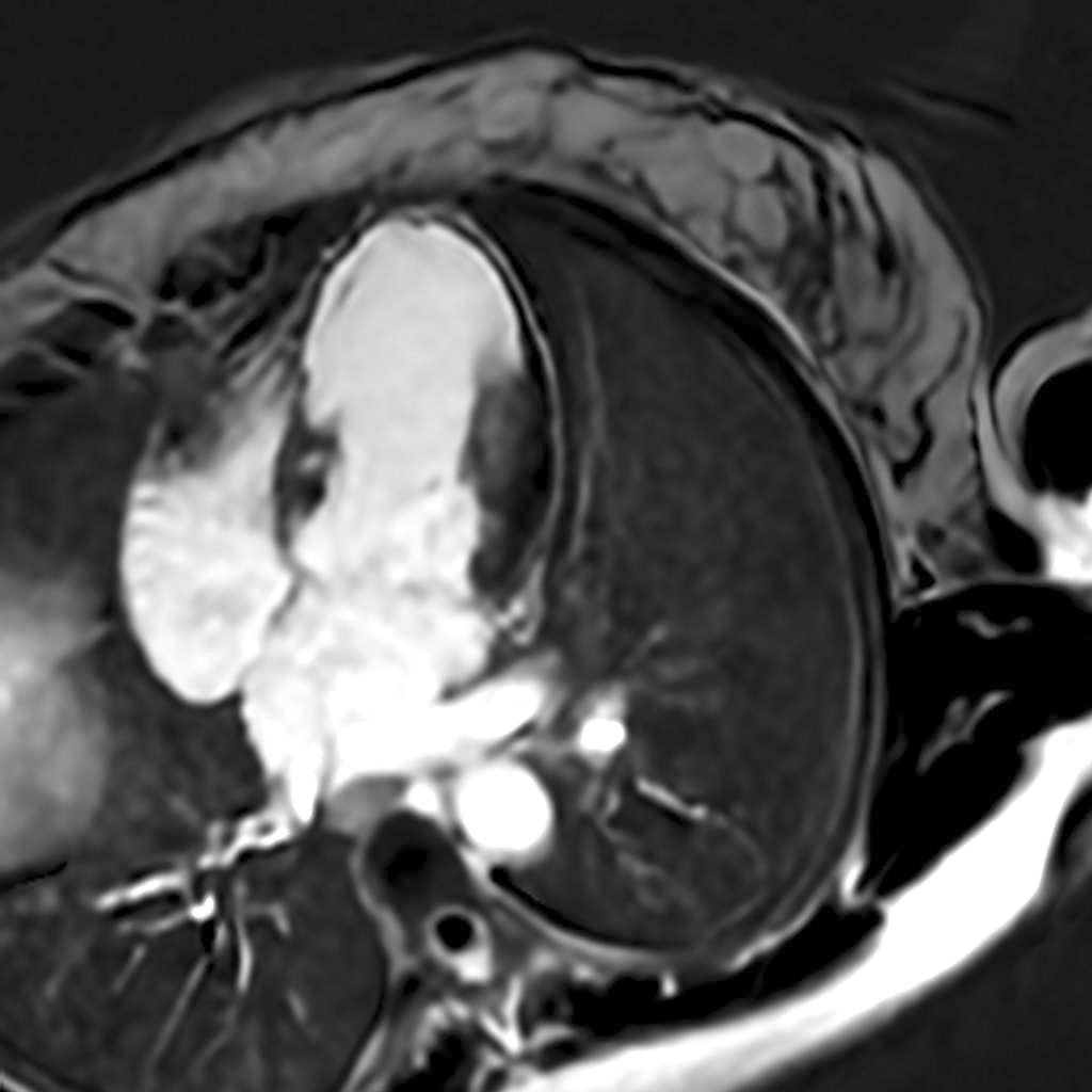
Cardiac MRI allows cardiologists to assess heart function and structure in remarkable detail. It can reveal pumping ability, chamber size, scarring, inflammation such as myocarditis, abnormal protein buildup like amyloid, and whether heart muscle is still viable, insights other imaging tests cannot provide.
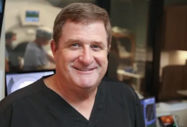
Dr. Richard Thompson, M.D., FAAC, FSCMR, medical director of advanced cardiac imaging at Health First, has performed more than 12,000 cardiac MRIs over his career. He specializes exclusively in cardiac MRI interpretation, with many cases requiring an hour or more of careful analysis.
“We’re often caring for patients with complex or advanced heart disease,” Thompson said. “This imaging gives us the highest level of detail when it matters most.”
What patients may not realize is just how precise a cardiac MRI can be. Working together, Thompson and the cardiac MRI technologists can identify areas of concern as small as the tip of a pencil on the heart muscle.
The GE Healthcare MRI systems at Health First are among the most advanced available, equipped with cutting-edge acceleration technology that allows for faster, highly reliable exams without compromising image quality.
For patients, the experience can feel intimidating at first. MRI machines produce loud knocking and thumping sounds as they capture detailed “slices” or images of the heart from multiple angles. Because the heart is constantly moving and patients are breathing, capturing crisp images requires speed and precision.
“It’s like trying to take a picture of a moving target,” Thompson said.
Patients are given earplugs and often music to make the experience more comfortable. They are also asked to hold their breath for short periods to reduce motion artifacts, subtle distortions that can affect image quality. Most cardiac MRIs require a safe contrast injected through an IV, which highlights different disease processes in the heart muscle.
“Patients usually feel a cool flush,” Chris Womersley, R.T (R) (MR)(ARRT), an MRI technologist at Health First, explained. “Unlike CT contrast, which often feels warm, cardiac MRI contrast typically feels cool.”
Some patients also undergo stress imaging. While many have had a treadmill stress test, medication can safely simulate stress on the heart for a more detailed evaluation.
As patients hear the MRI’s various noises and follow instructions for breath holds, Womersley and Thompson communicate constantly via text to fine-tune each view, ensuring that any concerns or additional images are addressed immediately.
“The heart is unique to each person, almost like a fingerprint,” Womersley said.
While Womersley follows a specific set of imaging slices, both he and Thompson consider the patient’s symptoms and the referring physician’s notes. That collaboration helps them not only identify potential areas of concern but also recognize when the heart is not showing signs of a suspected condition — clarity that can reassure patients.
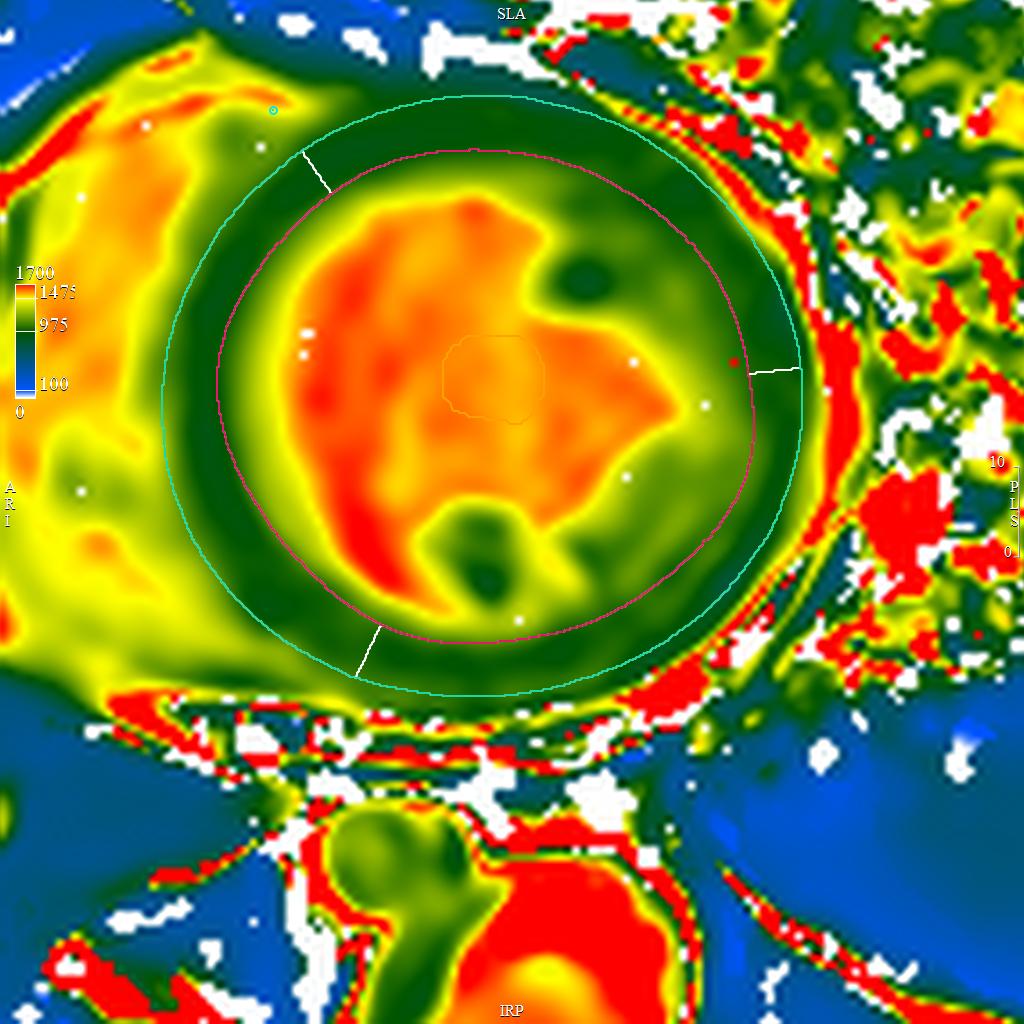
Womersley explained that the final images Thompson creates in real time look very different from typical MRI scans.
“We call them his ‘hurricane maps,’” he said with a laugh. “The swirling colors almost look like a weather radar, and they reveal incredible detail about the heart.”
“It’s very precise and technical,” Thompson said. “Cardiac MRI is a focused field, and even other physicians are sometimes surprised by how much goes into interpreting each study.”
That level of specialization is why physicians across Florida refer patients to Health First. The system performs roughly 1,000 cardiac MRIs each year, more than three times the number considered “high-level” at most centers, where 300 per year is already impressive.
Patients routinely travel from Jacksonville, Orlando, and Miami, with referrals coming from major health systems and Veterans Affairs facilities.
While many hospitals offer MRI, cardiac MRI is different. It requires specialized protocols, advanced software, and deep expertise.
“This is the gold standard,” Thompson said. “It allows us to analyze tissue characteristics such as fat, fluid, and scar tissue, something no other imaging test can do at this level.”
That clarity can directly influence life-altering decisions. One critical use is determining myocardial viability. If the heart muscle has died after a heart attack, bypass surgery may not help.
“If the tissue is dead and there’s a lot of it, there’s no point in bypassing it,” Thompson said. “Cardiac MRI helps us make those life-altering decisions.”
While a patient is being scanned, Thompson is already reviewing images using advanced software. He draws detailed contours around heart chambers and analyzes mapping sequences that measure tissue changes.
Each case is carefully reviewed to provide referring physicians with a precise assessment of the heart’s structure, function and tissue health.
Disease patterns often appear in specific regions of the heart. Cardiac MRI can show whether damage is in the apex, midsection, or base, helping physicians better understand the underlying cause. It can also identify inflammation, scarring, or clots.
When asked about artificial intelligence, Thompson describes it as a tool, not a replacement. Every image is still reviewed by a physician, and every finding is discussed with the referring doctor. From the MRI control room to the advanced software he uses, AI enhances accuracy and efficiency without replacing human expertise.
One of the most reassuring aspects for patients is how quickly results become available. In many cases, results appear in the MyChart app, powered by Epic, even before patients leave the hospital. Follow-up with the referring physician is still essential to review findings and determine next steps.
For some patients, cardiac MRI reveals reassuring news. For others, it identifies high-risk conditions that require intervention.
“Based on what I see on the images, a patient may be at higher risk for future cardiac events,” Thompson said.
“That’s why physicians refer them to us.” While he cannot predict exactly when something might happen, his insights help doctors determine whether a patient needs medication adjustments, a defibrillator, or more intensive treatment.
“That’s the power of cardiac MRI,” he said. “There aren’t many tools that can provide this level of guidance. This capability has transformed our cardiology practice and, most importantly, has given patients clarity and options they didn’t have before.”
For patients and families navigating uncertainty about heart health, the advanced cardiac MRI program at Health First offers more than just images. It provides clarity, guidance, and confidence, helping doctors make informed decisions and giving patients peace of mind.
From detecting tiny areas of concern to confirming when the heart is healthy, these scans bring answers when they matter most. Sometimes, what ends with a colorful map on a screen can make a life-changing difference.