What Is Lumbar Stenosis? Causes And Symptoms Explained
By Space Coast Daily // April 9, 2025

Lumbar stenosis is a common spinal condition that affects millions of people, particularly older adults. It occurs when the spinal canal located in the lower back experiences narrowing, putting pressure on the nerves that travel through the spine.
This compression can lead to pain, numbness, and weakness in the legs and lower body. Understanding the causes, symptoms, and treatment options is essential for managing this condition effectively.
This article explores lumbar stenosis in detail, covering its causes, symptoms, diagnostic methods, and treatment options.
Understanding Lumbar Stenosis
Lumbar stenosis is a condition that primarily affects the lower back, specifically the lumbar region of the spine. It occurs when the spinal canal, which houses the spinal cord and nerve roots, becomes narrowed. This narrowing can be caused by a variety of factors, including age-related degeneration, injuries, or congenital conditions. As the spine ages, the discs between vertebrae lose hydration and elasticity, which can lead to a reduction in space within the spinal canal. Over time, bone spurs and thickened ligaments can further contribute to the narrowing, putting pressure on the spinal cord and nerves.
There are two main types of lumbar stenosis, such as:
■ Central Stenosis: This refers to the narrowing of the spinal canal in the center of the lumbar spine, where the spinal cord is located. This type of stenosis can lead to direct compression of the spinal cord, causing symptoms such as lower back pain, numbness, weakness, or even difficulty walking. In severe cases, it can lead to more serious neurological issues, such as loss of bladder or bowel control.
■ Foraminal Stenosis: This occurs when the openings (foramina) through which the nerve roots exit the spine become compressed. This can result in radiating pain, often felt in the legs or feet, a condition known as sciatica. In addition to pain, patients may experience tingling, burning sensations, or weakness in the affected leg. The pain typically worsens with certain movements, such as standing for long periods or bending backward.
Indeed, the spine is made up of vertebrae, discs, and nerves that work together to support the body and facilitate movement. When the space within the spinal canal shrinks, it can compress the spinal cord or nerve roots, leading to discomfort and mobility issues.
While lumbar stenosis primarily involves the spine, persistent neurological symptoms may require evaluation by a brain specialist to rule out coexisting conditions like cauda equina syndrome or other spinal cord disorders. Early specialist consultation ensures comprehensive care and prevents permanent nerve damage.
Causes of Lumbar Stenosis
Several factors contribute to the development of lumbar stenosis. These include:
Age-Related Degeneration
As people age, natural wear and tear on the spine can lead to degenerative changes. The intervertebral discs, which act as cushions between the vertebrae, lose moisture and elasticity, causing them to shrink and become less effective at absorbing shock. This may result in a reduction of the space within the spinal canal, which can place pressure on the spinal cord and nerves.
Herniated Discs
A herniated or slipped disc can also contribute to lumbar stenosis. When the soft inner material of a disc bulges out through a tear in the tougher outer layer, it can protrude into the spinal canal. This protrusion can compress the nearby spinal nerves or the spinal cord itself, causing symptoms of lumbar stenosis.
Spinal Osteoarthritis
Osteoarthritis (OA) is a degenerative joint disease characterized by the breakdown of cartilage, the tissue that cushions the ends of bones. As the cartilage deteriorates, bones rub against each other, leading to pain, stiffness, and swelling. This condition can affect any joint, but it most commonly impacts weight-bearing joints, such as the hips, knees, and spine.
Congenital Conditions
Some individuals are born with a naturally narrow spinal canal, a condition known as congenital lumbar stenosis. This narrowness can cause insufficient space within the canal for the normal passage of the spinal cord and nerve roots. Although many people with congenital spinal stenosis may not experience symptoms during their early years, the condition can lead to problems as they age.
Spinal Injuries
Spinal injuries, whether caused by trauma or surgery, can also lead to spinal stenosis by causing structural changes to the spine. Accidents, such as car crashes or falls, can lead to fractures or dislocations of the vertebrae. These structural changes may push bone fragments or displaced vertebrae into the spinal canal, resulting in nerve compression. Similarly, surgeries performed on the spine, while often necessary for treating other conditions, can lead to complications such as scar tissue formation or misalignment of vertebrae, which can narrow the spinal canal\
Symptoms of Lumbar Stenosis
The symptoms of lumbar stenosis vary depending on the severity and location of nerve compression. As the spinal canal narrows, pressure on the nerves can lead to a range of discomforts that worsen over time.
Some common symptoms of lumbar stenosis include:
Lower Back Pain
A dull, persistent ache or stiffness in the lower back can be one of the earliest signs. This discomfort often intensifies after prolonged standing, walking, or physical activity. Some individuals experience relief when sitting or leaning forward, as this posture can temporarily reduce pressure on the compressed nerves.
Leg Pain and Numbness (Sciatica)
When nerves in the lumbar spine are compressed, sharp, burning, or electric-like pain can radiate down the buttocks and legs—a condition known as sciatica. Tingling or numbness may accompany the pain, typically affecting one or both legs. Symptoms can worsen with movement but may ease with rest.
Weakness in the Legs or Feet
As nerve compression progresses, muscle weakness can develop, particularly in the calves or feet. This may cause difficulty walking, frequent tripping, or a sensation of heaviness in the legs. In severe cases, foot drops or difficulty lifting the front of the foot can occur, increasing the risk of falls. Hence, early recognition of these symptoms is crucial for timely treatment and preventing further nerve damage.
Improved Symptoms with Sitting or Bending Forward
One of the hallmark signs of lumbar stenosis is symptom relief when sitting or bending forward. This occurs because flexing the spine slightly opens the spinal canal, reducing pressure on compressed nerves.
Many patients report that standing upright or walking for extended periods worsens their pain, while leaning on a shopping cart, sitting down, or crouching provides noticeable relief. This phenomenon, known as “neurogenic claudication,” distinguishes lumbar stenosis from other back conditions. However, prolonged sitting can eventually lead to stiffness, so alternating positions are often recommended for comfort.
Loss of Bladder or Bowel Control (In Severe Cases)
In rare but critical cases, severe nerve compression can lead to cauda equina syndrome, a medical emergency. This occurs when the bundle of nerves at the base of the spinal cord (cauda equina) is severely compressed, causing loss of bladder or bowel control, numbness in the saddle area (inner thighs, buttocks), and sudden leg weakness. As such, immediate surgical intervention is required to prevent permanent nerve damage and paralysis.
Diagnosing Lumbar Stenosis
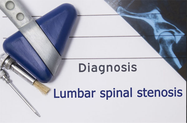
A comprehensive medical evaluation is essential for accurately diagnosing lumbar stenosis. The process typically includes:
Medical History and Physical Examination
A physician reviews the patient’s symptoms, medical history, and lifestyle factors. Physical tests, such as checking reflexes, muscle strength, and flexibility, can help assess nerve function and pinpoint areas of compression.
Imaging Tests
Many imaging tests are used to diagnose lumbar stenosis. For instance, x-rays serve as the initial imaging modality in evaluating lumbar stenosis, primarily revealing structural abnormalities of the vertebrae. These two-dimensional images can effectively detect bone spurs (osteophytes), vertebral fractures, spinal misalignment (spondylolisthesis), and degenerative changes associated with osteoarthritis. While x-rays don’t show soft tissues or nerves, they can provide valuable information about bony anatomy and spinal stability, helping clinicians determine if more advanced imaging is necessary.
Moreover, MRI has become the gold standard for diagnosing lumbar stenosis due to its unparalleled ability to visualize soft tissue structures. This non-invasive technology uses powerful magnets and radio waves to generate high-resolution, three-dimensional images of the spinal canal contents. MRIs can clearly show disc herniations, ligamentum flavum hypertrophy, facet joint degeneration, and the degree of nerve root compression. Detailed soft tissue visualization can help differentiate between central canal stenosis and foraminal narrowing, while also ruling out other conditions like tumors or infections that might mimic stenosis symptoms.
Lastly, CT scans offer exceptional bony detail through cross-sectional images reconstructed from multiple x-ray projections. When combined with myelography (CT myelogram), this modality can provide excellent visualization of bone and neural structures. CT is also particularly valuable for patients who can’t undergo MRI due to pacemakers or claustrophobia or when detailed assessment of bony anatomy is required before surgical intervention. The technology excels at revealing subtle fractures, facet joint arthritis, and calcifications that might contribute to stenosis.
Nerve Conduction Studies
Electromyography (EMG) measures electrical activity in muscles, while nerve conduction studies assess how well signals travel through nerves. These tests help identify the exact location and extent of nerve damage. Early and accurate diagnosis ensures appropriate treatment, preventing further deterioration and improving quality of life.
Treatment Options for Lumbar Stenosis
The treatment approach for lumbar stenosis depends on the severity of symptoms and their impact on daily life. Most cases begin with conservative, non-surgical methods before considering more invasive options.
Non-Surgical Treatments
Individuals diagnosed with lumbar stenosis can take advantage of several non-surgical treatments. These include:
■ Physical Therapy: A structured rehabilitation program focuses on strengthening core and back muscles, improving flexibility, and enhancing posture. Therapists may incorporate low-impact exercises, stretching, and aquatic therapy to reduce spinal pressure while maintaining mobility.
■ Medications: NSAIDs (Nonsteroidal Anti-Inflammatory Drugs) like ibuprofen can help reduce inflammation and pain. Oral steroids may be prescribed for short-term relief of severe inflammation. Also, epidural steroid injections can deliver anti-inflammatory medication directly to the affected nerve roots, providing weeks to months of pain relief.
■ Lifestyle Modifications: Weight loss reduces stress on the lumbar spine. Posture correction and ergonomic adjustments (e.g., supportive chairs, proper lifting techniques) can also minimize strain. Lastly, low-impact activities like swimming or cycling maintain fitness without worsening symptoms.
Surgical Interventions
When conservative treatments like physical therapy and medications fail to provide adequate relief after 3-6 months, or when patients experience worsening neurological symptoms (such as progressive weakness or loss of bladder control), surgical intervention becomes necessary. The two most common surgical approaches include:
■ Laminectomy (Decompression Surgery): This gold-standard procedure involves removing the lamina (the bony roof of the vertebral arch) to create more space in the spinal canal, immediately relieving pressure on compressed nerves. Surgeons may also remove bone spurs or thickened ligaments contributing to stenosis. The procedure also typically takes 1-2 hours, with most patients experiencing significant pain relief and improved mobility during recovery.
■ Spinal Fusion: Often performed concurrently with laminectomy when spinal instability exists, fusion permanently connects two or more vertebrae using bone grafts and metal hardware. While this stabilizes the spine and prevents painful motion, it slightly reduces flexibility. Newer minimally invasive techniques have shortened recovery times to about 3-6 months for both procedures.
The decision between these options depends on each patient’s specific anatomy, symptom severity, and overall health profile.
Preventing Lumbar Stenosis
While age-related spinal degeneration is inevitable, adopting proactive lifestyle measures can significantly slow stenosis progression and preserve spinal health. For instance, a targeted exercise regimen focusing on core and back muscle strengthening can help maintain proper spinal alignment and reduce mechanical stress on vertebral structures. Postural awareness is equally crucial—whether sitting at a desk, standing for prolonged periods, or lifting objects, proper body mechanics (engaging leg muscles rather than bending at the waist) can minimize unnecessary spinal compression.
Furthermore, smoking cessation plays a vital protective role, as nicotine’s vasoconstrictive effects can impair nutrient delivery to spinal discs, hastening their breakdown. Low-impact activities like yoga can enhance flexibility while improving circulation to spinal tissues, and regular walking can maintain joint mobility without excessive strain.
Early implementation of these preventive strategies and prompt attention to initial symptoms form the foundation for long-term spinal health. Such measures can delay stenosis progression and enhance overall musculoskeletal function, helping individuals maintain active lifestyles despite natural age-related changes.
Conclusion
Lumbar stenosis is a progressive spinal condition that can severely limit mobility and diminish quality of life if left untreated. Early diagnosis through proper medical evaluation and imaging is critical for implementing effective treatment strategies. While conservative approaches like physical therapy and medication often provide relief, surgical options may be necessary in advanced cases. By keeping the information mentioned above in mind, patients can prevent permanent nerve damage and the overall risk of lumbar stenosis.